
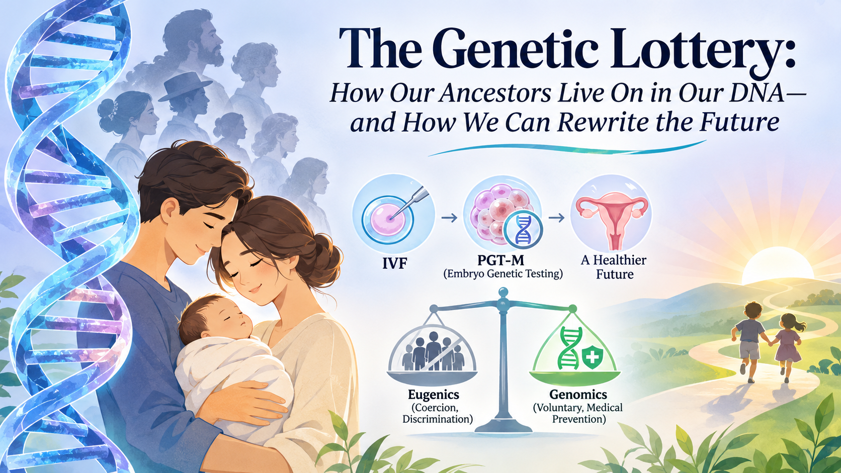
Our DNA carries fragments of biological history, including genetic variations that can shape the health of generations to come. But what happens when an inherited mutation threatens the life of a future child? Advances in reproductive genetics are giving families new possibilities. They are also forcing humanity to reconsider an important question: how do we use the power to influence genetic inheritance without repeating the mistakes of the past?
Key takeaways
Inheritance is more complicated than a family tree: Genetic disorders can follow autosomal dominant, autosomal recessive, X-linked and other inheritance mechanisms. Not every genetic condition follows a predictable Mendelian pattern.
Genetic risk can sometimes be identified before pregnancy: Genetic testing, carrier screening and genetic counselling can help prospective parents understand reproductive risks.
PGT-M offers an additional reproductive option: Preimplantation genetic testing for monogenic conditions can identify embryos carrying specific familial disease-associated variants before embryo transfer through IVF.
Testing has limitations: PGT-M does not screen every possible genetic disorder, guarantee a healthy child or eliminate the need for appropriate prenatal care.
Genetic medicine must learn from history: Modern reproductive genetics must protect reproductive autonomy, informed consent and the dignity of people living with disabilities.
The objective is informed choice, not genetic perfection: Preventing a serious inherited disease and attempting to engineer supposedly superior human beings are fundamentally different objectives, but the ethical boundaries require continuous examination.
1. The blueprint of life: Understanding genetic inheritance
Every human being inherits genetic information from their biological parents. This information is stored in DNA (deoxyribonucleic acid), the molecule that carries the instructions necessary for biological development and functioning.
Human cells generally contain 46 chromosomes arranged in 23 pairs. One chromosome from each pair is inherited from the biological mother and the other from the biological father.
Within these chromosomes are approximately 20,000 protein-coding genes, alongside extensive non-coding DNA that also contributes to gene regulation and other biological functions.
A change in the DNA sequence of a gene is called a genetic variant. Some variants have no known health consequences. Others may influence disease susceptibility, while certain pathogenic variants can directly cause inherited disorders.
An important distinction is that not all genetic disorders are inherited from parents. Some arise spontaneously, known as de novo variants, during the formation of reproductive cells or early embryonic development.
According to the US Centers for Disease Control and Prevention (CDC), genetic disorders include single-gene conditions, chromosomal abnormalities and complex conditions involving multiple genetic and environmental factors.
The central principle: A person's genetic inheritance can influence their health, but it does not independently determine every aspect of their future health.
Scientific reference: CDC – Genetic Disorders
2. How genetic diseases travel through generations
Understanding inheritance patterns is essential when estimating the likelihood that a child may inherit a genetic condition.
The three commonly discussed Mendelian inheritance patterns are autosomal dominant, autosomal recessive and X-linked inheritance.
A. Autosomal dominant inheritance
Autosomal dominant disorders occur when a pathogenic variant in one of the two copies of a gene is sufficient to cause or predispose an individual to a condition.
The term autosomal means that the affected gene is located on one of the 22 pairs of non-sex chromosomes.
Consider a parent carrying one disease-associated variant and one unaffected copy of the gene.
If the condition follows a conventional autosomal dominant inheritance pattern, each pregnancy has a:
50%
Probability of transmitting the variant to each child
This probability applies independently to every pregnancy. It does not mean that exactly half of all children in a family will inherit it.
Examples include:
Huntington disease.
Marfan syndrome.
Some forms of familial hypercholesterolemia.
An important qualification is that inheriting a pathogenic variant does not always mean that a person will develop the condition. Some variants demonstrate incomplete penetrance, while others produce different degrees of disease severity.
B. Autosomal recessive inheritance
Autosomal recessive disorders generally require pathogenic variants in both copies of a particular gene.
A person who has one pathogenic variant and one functioning copy is often called a carrier. Carriers of many recessive conditions do not develop the associated disease, although this is not universally true for every genetic condition.
Consider a couple in which both biological parents carry a pathogenic variant in the same disease-associated gene.
For each pregnancy, the possible outcomes are:
The 25–50–25 inheritance model
25%
Unaffected, non-carrier
50%
Unaffected carrier
25%
Affected
Illustrative probabilities for two carriers of pathogenic variants in the same autosomal recessive gene, assuming the relevant variants cause disease when inherited together.
Examples include:
Sickle cell disease.
Cystic fibrosis.
Spinal muscular atrophy (SMA).
Tay-Sachs disease.
This pattern is particularly important in reproductive genetics because both parents may be completely healthy while carrying disease-associated variants.
C. X-linked inheritance
X-linked disorders arise from pathogenic variants in genes located on the X chromosome.
Typically, females have two X chromosomes, while males have one X and one Y chromosome.
Because males generally have only one copy of most X-linked genes, a pathogenic variant in that copy can have a greater likelihood of producing disease.
In conventional X-linked recessive inheritance, a mother carrying a pathogenic variant has a 50% probability of transmitting that variant in each pregnancy. A son who inherits it may be affected, while a daughter who inherits it may be a carrier, although clinical manifestations in females are possible.
Examples include:
Duchenne muscular dystrophy.
Haemophilia A.
Some other X-linked neurological and metabolic disorders.
An important scientific distinction: Fragile X syndrome
Fragile X syndrome is frequently misrepresented as a conventional X-linked recessive condition.
It is associated with an expansion of CGG trinucleotide repeats in the FMR1 gene on the X chromosome.
The number of repeats matters:
FMR1 repeat category
Approximate repeat count
Intermediate
45–54
Premutation
55–200
Full mutation
More than 200
A full mutation is typically associated with abnormal methylation and reduced or absent production of the FMRP protein, which is important for normal brain development.
Premutation carriers can also face health risks, including fragile X-associated tremor/ataxia syndrome (FXTAS) and fragile X-associated primary ovarian insufficiency (FXPOI).
The risk of expansion from a maternal premutation to a full mutation depends partly on repeat size and other sequence characteristics, including AGG interruptions.
This is why genetic counselling for Fragile X requires more than simply identifying whether a parent carries an altered gene.
Primary clinical reference: GeneReviews, FMR1 Disorders, updated May 2024.
3. Beyond inheritance patterns: Why genetics is not always predictable
The three inheritance patterns above are useful starting points, but they do not explain every genetic disease.
Several additional mechanisms influence genetic inheritance and disease development:
De novo variants: Genetic changes that arise for the first time in an individual rather than being inherited from a parent.
Mitochondrial inheritance: Certain conditions arise from variants in mitochondrial DNA, which is predominantly transmitted through the maternal line.
Genomic imprinting: The expression of certain genes depends on whether they were inherited from the mother or father.
Repeat expansion disorders: Certain DNA sequences become abnormally expanded across generations, as seen in Fragile X and Huntington disease.
Multifactorial inheritance: Multiple genetic variations interact with environmental and lifestyle factors to influence disease risk.
These distinctions matter because the reproductive strategy appropriate for one disorder may not be suitable for another.
A genetic diagnosis must therefore establish the actual pathogenic variant, its inheritance mechanism and its clinical significance before reproductive decisions are made.
4. Breaking the chain: How PGT-M is changing reproductive medicine
For families carrying a serious inherited genetic condition, the possibility of transmitting that condition to a future child can be an emotionally difficult concern.
Traditionally, couples could pursue natural conception and consider prenatal diagnostic testing during pregnancy. Depending on the findings, they would then face decisions about pregnancy management.
Advances in assisted reproductive technology have introduced another option: Preimplantation Genetic Testing for Monogenic Conditions (PGT-M).
PGT-M is a specialized genetic testing procedure performed as part of an IVF treatment cycle. It helps identify embryos carrying specific disease-associated genetic variants before an embryo is selected for transfer into the uterus.
It is important to understand that PGT-M is not a standalone treatment or a guarantee of a disease-free child. It is a reproductive risk-reduction strategy.
How does PGT-M work?
The process generally involves the following stages.
Step 1: Genetic evaluation of prospective parents
Before IVF begins, the clinical team establishes the family's genetic diagnosis.
This may involve:
Reviewing family medical history.
Confirming the pathogenic variant in an affected family member or prospective parent.
Determining the inheritance pattern.
Assessing whether PGT-M is technically feasible.
In some cases, samples from relatives are needed to establish genetic linkage.
Step 2: In vitro fertilization
The prospective mother undergoes ovarian stimulation to produce multiple mature eggs.
The eggs are retrieved and fertilized with sperm in the laboratory, commonly using intracytoplasmic sperm injection (ICSI).
The resulting embryos are cultured under controlled laboratory conditions.
Step 3: Embryo development and biopsy
Embryos are generally cultured until the blastocyst stage, usually around day five or six.
At this stage, a small number of cells are removed from the trophectoderm, the outer cell layer that primarily contributes to the placenta.
The biopsy is sent for genetic analysis. The embryo is usually cryopreserved while testing is completed.
Step 4: Genetic analysis
The laboratory analyses the DNA from the biopsy to determine whether the embryo carries the specific familial genetic variant.
Two important approaches are:
Direct variant analysis: Identifies the known pathogenic variant itself.
Linkage analysis: Examines nearby genetic markers that are inherited alongside the disease-associated variant.
These methods may be used together to improve diagnostic reliability.
Because the amount of DNA obtained from an embryo biopsy is very small, DNA amplification is required before analysis.
Depending on the laboratory's methodology, amplification may be targeted, multiplex or whole-genome amplification.
Whole-genome amplification is therefore one possible laboratory technique, not a universal requirement for every PGT-M test.
Step 5: Embryo selection and transfer
The genetic findings are interpreted in the context of the specific condition and inheritance pattern.
Embryos may be classified as affected, unaffected carriers or unaffected non-carriers, depending on the disorder being investigated.
An embryo considered suitable for transfer may then be selected in consultation with the prospective parents and their medical team.
Importantly, some couples may choose to transfer an unaffected carrier embryo, especially in autosomal recessive conditions, because being a carrier does not ordinarily mean having the disease.
The final decision requires condition-specific counselling.
Which diseases can PGT-M help prevent?
The American Society for Reproductive Medicine (ASRM) identifies several conditions for which PGT-M may be considered.
Condition
Associated gene
Sickle cell disease
HBB
Spinal muscular atrophy
SMN1
Tay-Sachs disease
HEXA
Cystic fibrosis
CFTR
Huntington disease
HTT
Hereditary breast and ovarian cancer syndrome
BRCA1 / BRCA2
The clinical implications differ considerably between these conditions. Some are severe childhood-onset disorders, whereas others involve adult-onset disease, variable penetrance or different levels of clinical severity.
PGT-M suitability must be evaluated individually.
Primary source: ASRM Practice Committee, Indications and Management of Preimplantation Genetic Testing for Monogenic Conditions, 2023.
Read the complete clinical committee opinion
5. The science has limitations: What PGT-M cannot promise
The development of PGT-M is a major advancement, but responsible medical communication requires discussing its limitations just as clearly as its benefits.
There are several reasons why PGT-M should not be interpreted as a guarantee of a healthy child.
A. It tests for specific genetic conditions
PGT-M is designed to investigate particular known genetic variants associated with a familial disorder.
It does not automatically screen an embryo for every genetic disease, developmental condition or future health risk.
An embryo that tests negative for the specific variant under investigation may still carry other genetic abnormalities that have not been tested.
B. Diagnostic errors are possible
PGT-M involves analysing a very small amount of embryonic DNA. Technical challenges can affect the results.
Some important limitations include:
Allele dropout: One of the two gene copies fails to amplify or is not detected.
Contamination: Extraneous DNA affects the test result.
Recombination: Genetic material is exchanged between chromosomes, potentially complicating linkage analysis.
Insufficient DNA quality: The available sample does not produce a reliable result.
Modern laboratory techniques are designed to reduce these risks, but they cannot eliminate them entirely.
C. Not every embryo will be suitable for transfer
The number of embryos available for transfer depends on several factors, including maternal age, ovarian reserve, fertilisation rates, embryo development and the inheritance pattern of the condition.
For example, if both parents carry a pathogenic variant for an autosomal recessive disorder, approximately 25% of embryos are expected to inherit both variants under the conventional inheritance model.
However, this probability is not a prediction of the number of transferable embryos. Other clinical and laboratory factors influence the final outcome.
D. Prenatal confirmation remains important
The American College of Obstetricians and Gynecologists (ACOG) and ASRM recommend offering prenatal diagnostic confirmation following pregnancies achieved using PGT-M.
Depending on the circumstances, this may involve:
Chorionic villus sampling (CVS).
Amniocentesis.
These procedures allow diagnostic testing during pregnancy and can help confirm the earlier embryo-testing findings.
PGT-M also does not replace routine prenatal screening and assessment for unrelated fetal conditions.
Clinical references:
6. A darker chapter in scientific history: The rise of eugenics
The ability to examine genetic information and influence reproductive outcomes introduces an important ethical question:
If science gives us the ability to influence which genetic characteristics are passed to future generations, who decides which characteristics should be selected?
This question cannot be answered without understanding the history of eugenics.
What was eugenics?
The term eugenics was coined by English statistician and scientist Francis Galton in 1883.
Galton proposed that human populations could be improved through the selective reproduction of individuals whom he considered genetically desirable.
His ideas were rooted in deeply prejudiced assumptions about heredity, intelligence, social class and race.
Eugenics eventually developed into an international movement that attempted to classify people as genetically superior or inferior.
Its proponents believed that undesirable characteristics could be eliminated from society by controlling reproduction.
These ideas were scientifically flawed and ethically disastrous.
The United States: When pseudoscience became public policy
The American eugenics movement demonstrates how genetic determinism can become a tool of institutional discrimination.
Indiana, 1907
Indiana became the first US state to enact a compulsory sterilisation law in 1907.
The law authorised sterilisation of certain individuals held in state institutions, based on discriminatory judgments about their supposed hereditary fitness.
The policy was subsequently challenged and later replaced by further legislation.
The Indiana sterilisation programme continued until 1974. Historical estimates indicate that approximately 2,500 people were sterilised in the state under the broader programme.
The wider United States
The movement spread across the country.
By the 1930s, more than 30 states had enacted eugenic sterilisation laws.
The National Human Genome Research Institute (NHGRI) reports that more than 60,000 people were forcibly sterilised under these policies before the programmes were largely dismantled.
The affected populations included people with disabilities, poor communities, racial minorities and individuals labelled socially undesirable.
The Immigration Act of 1924
Eugenic thinking also influenced American immigration policy.
The Johnson-Reed Act of 1924 established restrictive national-origin quotas that sharply reduced immigration from Southern and Eastern Europe and effectively excluded most Asian immigrants.
These policies reflected racial and ethnic hierarchies rather than scientifically defensible assessments of human worth.
The consequences of these policies extended far beyond medicine. They affected families, communities, reproductive freedom and fundamental human rights.
Sources:
7. Modern genomics versus eugenics: Where is the ethical boundary?
It is tempting to conclude that modern genetic testing and historical eugenics have nothing in common.
That would be an oversimplification.
Both involve understanding human heredity and, in certain circumstances, making decisions about which genetic characteristics should be passed to future generations.
However, their underlying principles and practices can be fundamentally different.
Historical eugenics
Ethical clinical genomics
State-imposed reproductive control
Voluntary reproductive decisions
Racial and social hierarchies
Evidence-based clinical assessment
Forced sterilisation
Informed consent
Discrimination against people with disabilities
Respect for disability rights and dignity
Claims of genetic superiority
Reduction of specific medical risks
Population-level control
Individual and family-centred counselling
Modern clinical genomics should be based on patient autonomy, informed consent, scientific evidence and non-discrimination.
However, the ethical discussion does not end simply because testing is voluntary.
There are still difficult questions.
Could embryo selection gradually shift from preventing serious disease to selecting preferred physical characteristics?
Could expensive reproductive technologies create a society in which genetic advantages become accessible primarily to wealthy families?
Could people living with disabilities feel that society considers their existence undesirable?
Who should determine whether a particular genetic condition is sufficiently serious to justify embryo selection?
These are not merely hypothetical philosophical concerns. They are active subjects of debate in reproductive medicine and bioethics.
The NHGRI explicitly recognises the need to confront the historical misuse of genetics and the risks of contemporary scientific racism.
One particularly important distinction is between testing for a well-established pathogenic variant responsible for a serious inherited disease and attempting to predict complex characteristics such as intelligence, personality or social behaviour.
The latter involve many genetic and environmental influences and cannot be reduced to simple genetic selection.
The central ethical principle should be this: genetic technology must expand reproductive freedom, not establish a hierarchy of human worth.
Primary source: NHGRI – Eugenics and Scientific Racism
8. Practical implications: What prospective parents should know
For couples with a family history of inherited disease, understanding genetic risk before pregnancy can be valuable.
The following steps can help them make informed reproductive decisions.
Step 1: Understand your family history
Document known inherited conditions in both biological families.
Pay particular attention to:
Unexplained infant or childhood deaths.
Known inherited neurological disorders.
Recurrent miscarriages where a genetic cause is suspected.
Family members diagnosed with metabolic disorders.
Known carriers of conditions such as thalassaemia or sickle cell disease.
Family history can help identify individuals who may benefit from genetic evaluation, although the absence of a family history does not eliminate genetic risk.
Step 2: Consider appropriate genetic testing
Depending on the clinical circumstances, a specialist may recommend:
Carrier screening.
Diagnostic genetic testing.
Targeted testing for a known familial variant.
Chromosomal testing.
Genetic counselling.
For example, in India, carrier screening for haemoglobinopathies such as beta-thalassaemia is particularly relevant in populations where these conditions are prevalent.
Step 3: Consult a reproductive genetics specialist
If a pathogenic variant is identified, a genetic counsellor can help interpret its significance.
The discussion should cover:
Probability of transmission.
Expected disease severity and variability.
Available reproductive options.
Whether PGT-M is technically feasible.
Financial and emotional considerations.
Step 4: Understand all reproductive options
PGT-M is not the only possible approach.
Depending on individual circumstances, couples may consider:
Natural conception with prenatal diagnostic testing.
IVF with PGT-M.
Use of donor eggs or sperm.
Embryo donation.
Adoption.
Choosing not to pursue further reproductive intervention.
There is no universally correct decision. The appropriate choice depends on personal values, medical circumstances, financial resources and informed consent.
9. The future of reproductive genetics
Genomics is progressing beyond the identification of individual disease-causing variants.
Several emerging areas could influence reproductive medicine in the coming decades.
A. Next-generation sequencing
Advances in DNA sequencing have improved the ability to identify genetic variants associated with inherited conditions.
These technologies are contributing to more precise diagnosis and increasingly sophisticated reproductive genetic testing.
B. Improved embryo testing
Research into more accurate embryo biopsy methods, genetic amplification and molecular analysis may help reduce diagnostic uncertainty.
However, improved analytical technology does not automatically establish better pregnancy or live-birth outcomes.
C. Polygenic embryo screening
Unlike PGT-M, which targets specific monogenic disorders, polygenic embryo screening attempts to estimate the relative genetic risk of complex conditions using many genetic variants.
This field remains controversial.
Its predictive accuracy, clinical usefulness, transferability across different ancestry groups and ethical implications require careful evaluation.
It should not be confused with established clinical testing for a known familial pathogenic variant.
D. Gene editing
Technologies such as CRISPR-Cas systems have transformed molecular biology by allowing scientists to modify DNA sequences.
However, editing the genomes of human embryos intended for reproduction raises significant safety, ethical and regulatory concerns.
Heritable genome editing could introduce unintended genetic changes that are transmitted to subsequent generations.
It is important to distinguish laboratory research from an established and accepted reproductive treatment.
The future of reproductive genetics will therefore depend not only on scientific innovation but also on responsible regulation, transparent clinical evidence and public participation in ethical decision-making.
10. Conclusion: Our genes tell a story, but they do not have to write the entire future
Human inheritance is a remarkable biological process. Every generation carries genetic information from generations before it, including variations that can influence health and disease.
For families affected by serious inherited conditions, modern genetic medicine offers opportunities that were previously unavailable.
Carrier screening, genetic counselling, prenatal diagnosis and PGT-M have expanded the reproductive choices available to prospective parents.
But scientific progress must be accompanied by ethical responsibility.
The objective of reproductive genetics should not be to create a supposedly perfect human population or eliminate genetic diversity.
It should be to help individuals understand their reproductive risks, make informed decisions and access appropriate medical care without discrimination.
Our ancestors may have contributed the genetic material that makes us who we are. Modern science is giving us better ways to understand that inheritance. The responsibility is to use this knowledge with compassion, evidence and respect for human dignity.
Medical disclaimer
This article is intended for educational purposes and does not constitute individualised medical advice. Genetic risks, inheritance probabilities and the suitability of PGT-M depend on the specific condition, genetic variant and family circumstances. Individuals considering reproductive genetic testing should consult a qualified clinical geneticist, genetic counsellor and reproductive medicine specialist.
References and scientific literature
The following sources were consulted in preparing this article. Clinical recommendations and historical claims have been linked to the relevant primary or authoritative references.
Clinical genetics and inheritance
1. CDC. (2024). Genetic Disorders – Genomics and Your Health. Provides an overview of genetic disorders and Mendelian inheritance patterns.
2. Hunter, J. E., Berry-Kravis, E., Hipp, H., & Todd, P. K. (2024 update). FMR1 Disorders – GeneReviews, NCBI Bookshelf. Clinical reference covering Fragile X syndrome, repeat expansions, inheritance and genetic counselling.
Reproductive medicine and PGT-M
3. American Society for Reproductive Medicine Practice Committee. (2023). Indications and Management of Preimplantation Genetic Testing for Monogenic Conditions: A Committee Opinion. Fertility and Sterility, 120, 61–71.
4. American College of Obstetricians and Gynecologists. (2020). Preimplantation Genetic Testing. Clinical guidance concerning PGT and prenatal diagnostic confirmation.
History and bioethics
5. National Human Genome Research Institute. Eugenics and Scientific Racism. Historical and contemporary examination of eugenics and the misuse of genetic science.
6. National Human Genome Research Institute. Eugenics: Its Origin and Development (1883–Present). Historical timeline documenting Galton, American sterilisation policies and the development of the eugenics movement.
7. Indiana Historical Bureau. 1907 Indiana Eugenics Law. Historical record and discussion of the Indiana sterilisation programme.
8. US National Archives. Immigration from Asia and the Pacific, 1870s to 1950s. Historical documentation of restrictive US immigration legislation.
Research scope: This is a focused narrative literature review using authoritative clinical guidance, medical reference literature and historical institutional sources. It is not a systematic review or meta-analysis. The clinical guidance cited here includes the ASRM 2023 committee opinion and ACOG 2020 committee opinion; individual clinical practices and regulations may differ by country.